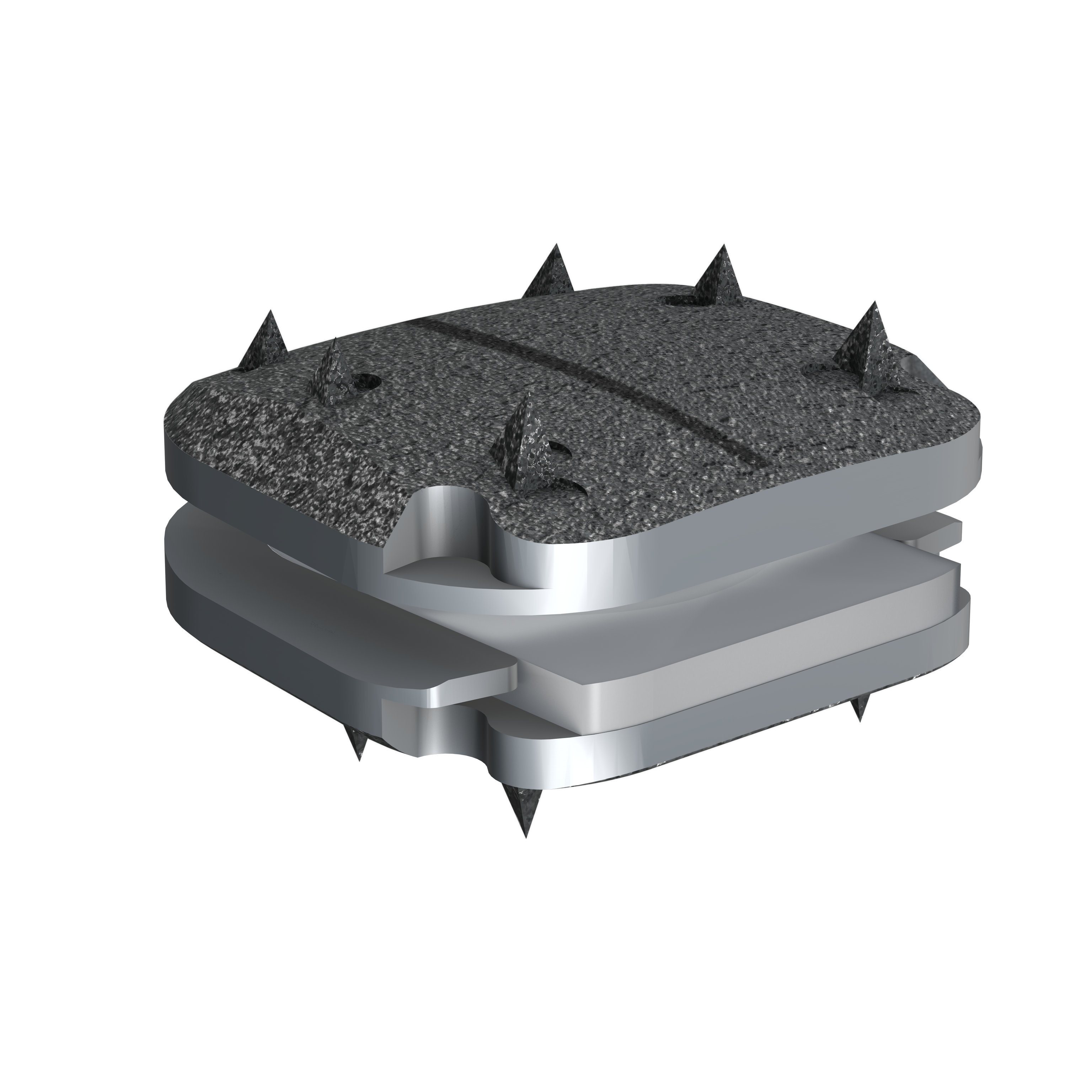
Image shows Isomorphic view of prodisc C Vivo.
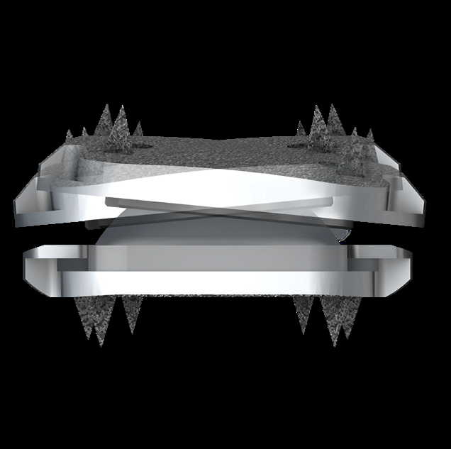
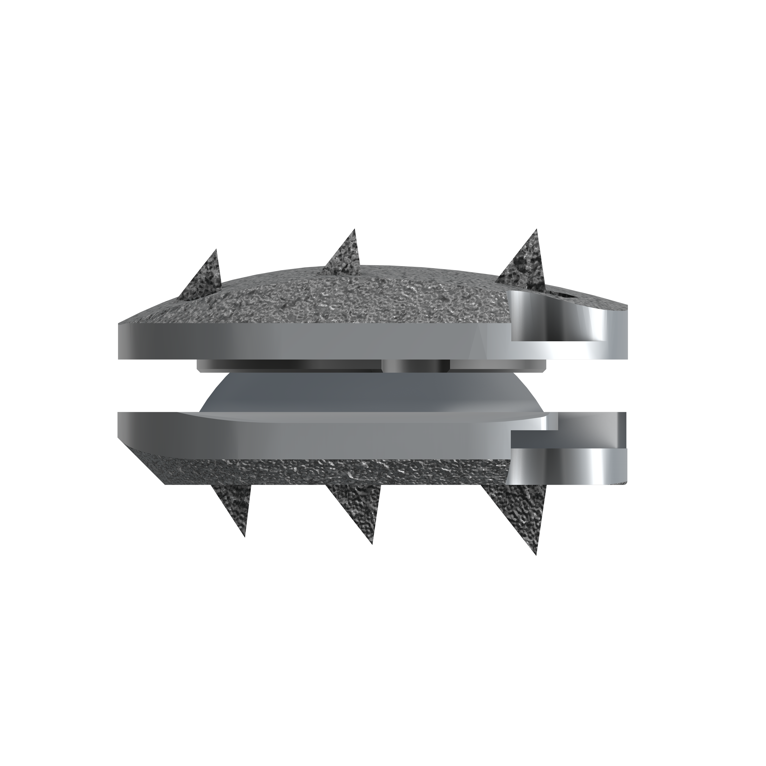
Image shows prodisc C Vivo lateral bending.
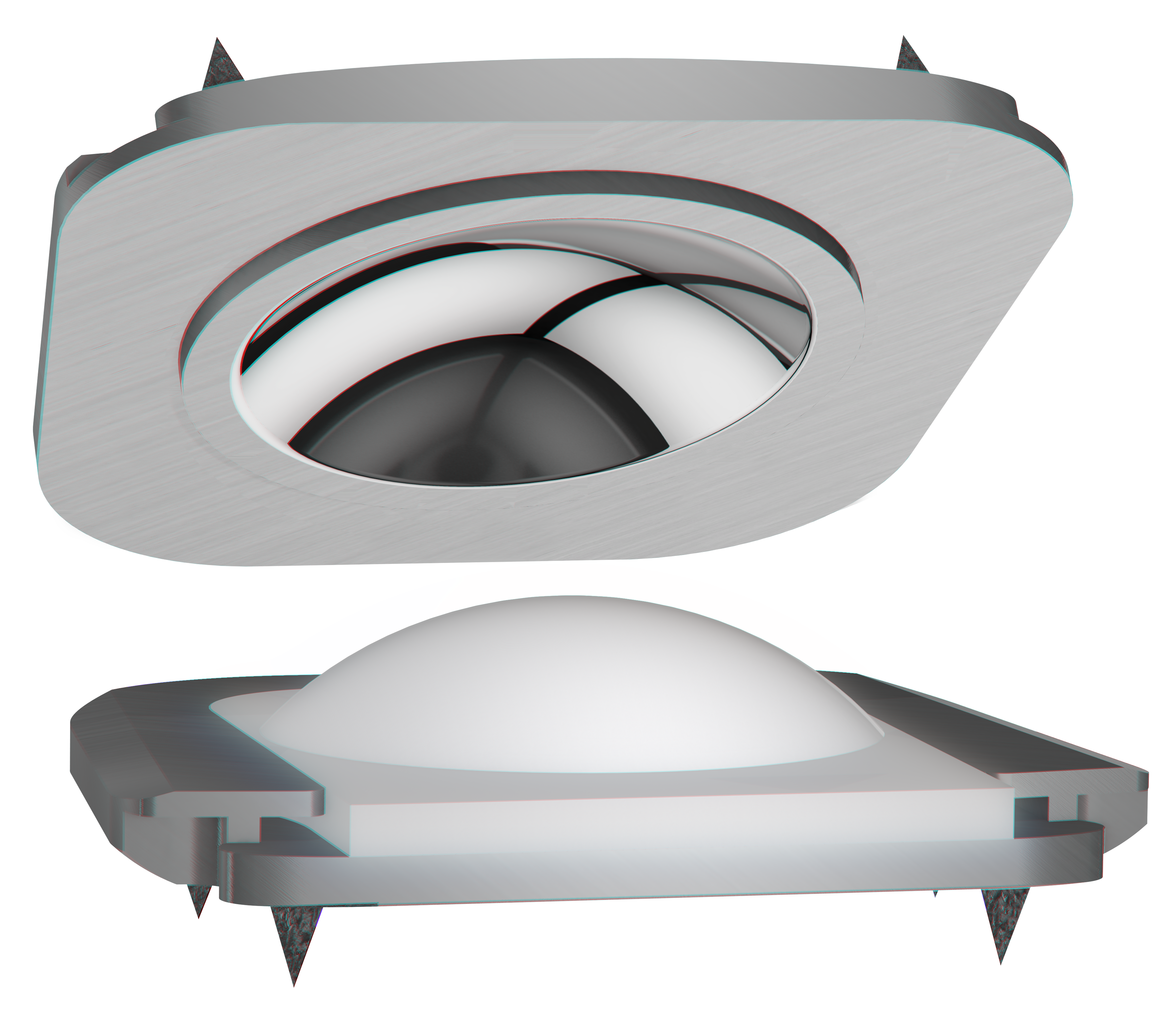
Image of ball and socket feature of the Vivo implant.
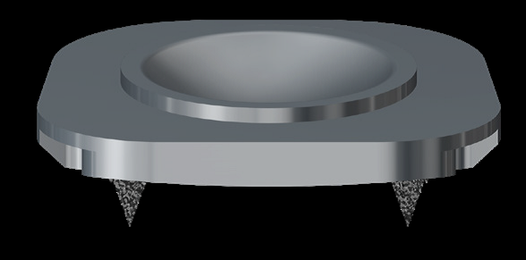
Image shows open view of superior endplate for prodis C Vivo.
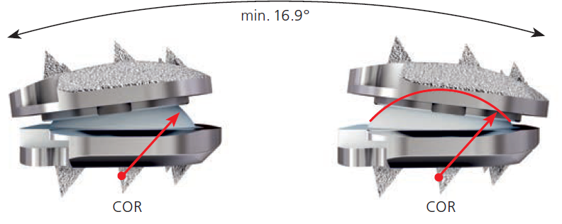
Image shows flexion-extension view of prodis C Vivo.
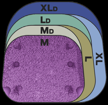
Image shows footprints available for prodisc C Vivo.
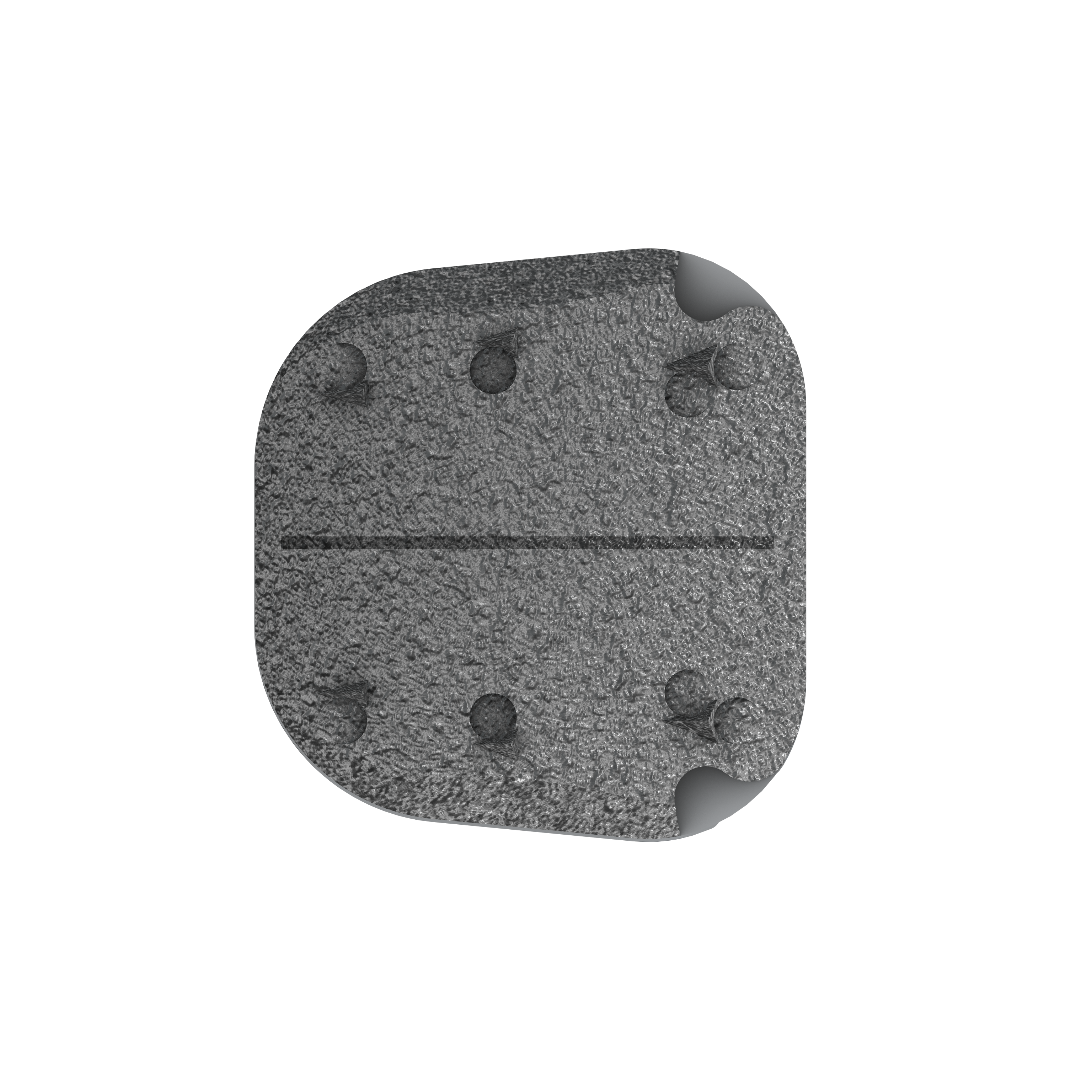
Overhead image of prodisc C Vivo showing the boney ongrowth surface
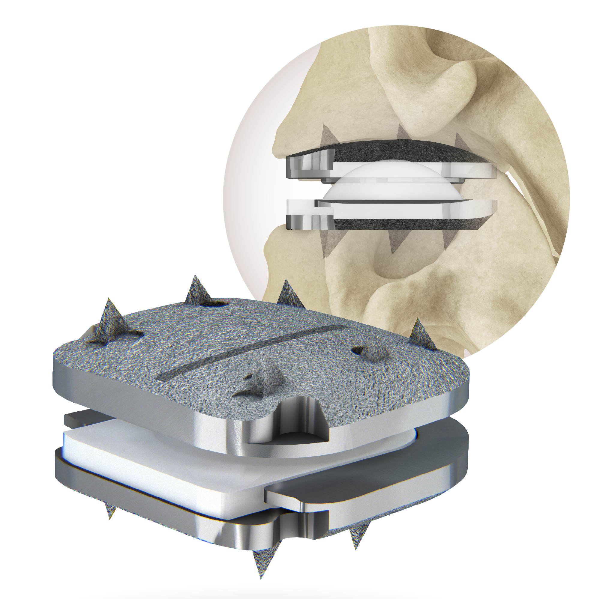
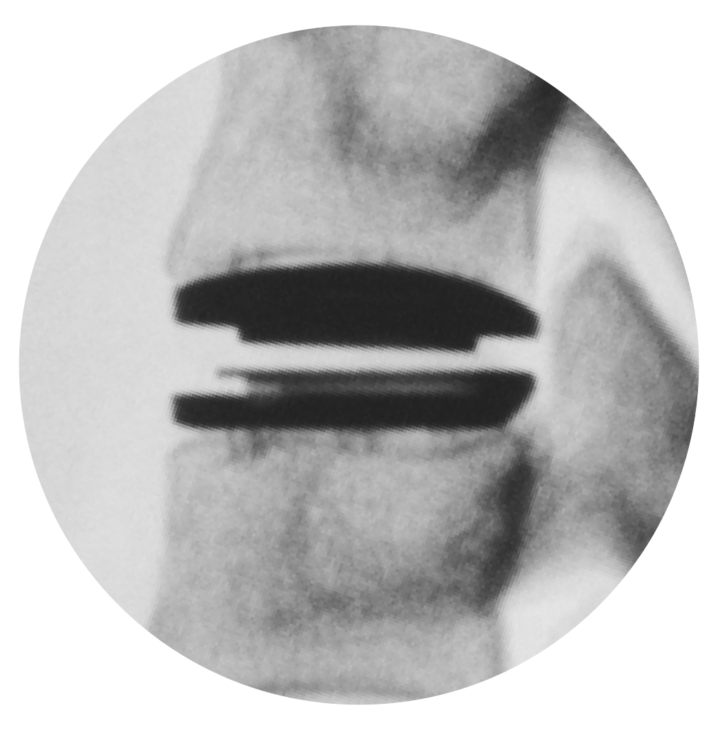
Image of prodisc C Vivo and implanted positioning.
Available Search Tags

Watch this short primer on Centinel Spine and its unique and extraordinary place as a catalyst of change in the spine industry—with pioneering technologies and a clinical history that have led to successes ranging from PGA champions to a growing list of surgeon-patients.