* The device system is designed for use with autograft bone and/or allogenic bone graft composed of cancellous and/or corticocancellous bone graft, to facilitate fusion.

No-Profile® Anterior Lumbar Integrated Interbody System
With Titanium-Enhanced PEEK Technology


Sixth generation STALIF® technology, STALIF M-Ti™ incorporates a novel, circumferential, titanium surface, PEEK Integrated Interbody™ design that is raising the Gold Standard of lumbar interbody fusion.


SEE HOW:

Optimized Surface Technology ↓
Integrally PEEK ↓
Inherently STALIF®


Top of the Page ↑
Optimized Surface Technology.1
Raising the Standard with


The Ti-ACTIVE™ surface provides a three-dimensional texturized surface that is microporous with hydrophilic properties, to facilitate human mesenchymal stem cell (hMSC) attachment and proliferation as demonstrated by pre-clinical testing performed on representative PEEK coupons with Ti-ACTIVE surface.2

During manufacturing, commercially pure titanium (CP Ti) is applied to the PEEK cage by a plasma spray to create our Ti-ACTIVE devices. The unique CP Ti surface system utilizes a robotic applicator, enabling the creation of a high-quality, reproducible titanium surface.3 Ti-ACTIVE was tested for mechanical integrity and the results exceeded the FDA requirements for shear and tensile strength for metallic spray surfaces.4 


1 Lebl DR. Mechanical performance of cervical TDRs in vivo. Spine (Phila, PA 1976) 2012 Dec 15; 37(26): 2151-60.
2 Yoon BJV, Cammisa FP, Abjornson C. Polishing methods less a factor in cell adhesion than surface characteristics of titanium plasma spray surfaces on PEEK. Paper presented at Lumbar Spine Research Society Meeting, 2015 April, Chicago, IL.
3 Centinel Spine Report VAL-2013-043.
4 Centinel Spine Report CD12-03-07-020.


Learn More About Ti-ACTIVE™ Technology  

Texturized Titanium Surface1

SEM image (2,000x magnification) of the surface of a standard PEEK implant (left) and a titanium-applied PEEK implant (right). Unlike standard PEEK, the PEEK with titanium surface provides a 3-D microporous environment conducive to cell attachment and cell migration, seen by cytoplasmic expansions of the cultured cells, as highlighted in purple.
Ti-ACTIVE™ PEEK Surface of STALIF M-Ti™

Cell Proliferation

SEM image (2,000x magnification) showing cellular interaction allowing human mesenchymal stem cells (hMSCs) to communicate when receptors and markers bind on a Ti-ACTIVE™ surface. Cell to cell interactions are vital in the promotion of cell adhesion and later the function of the cell to differentiate and proliferate, as highlighted in yellow. Ti-ACTIVE™ offers an optimized surface topography enabling cellular migration, cellular interaction, and cellular attachment to the surface as observed on representative STALIF® coupons with Ti-ACTIVE™ surface.2

Top of the Page ↑
STALIF M-Ti™ A/P View
STALIF M-Ti™ Lateral View

Radiolucent Advantage

The STALIF M-Ti™ device maintains the radiolucent advantage of PEEK, making it easier to assess post-operative development of fusion mass during follow-up.7 The Ti-ACTIVE™ surface allows for radiographic visualization of the graft-endplate contact. Images show radiographs of STALIF M-Ti™ in Anterior/Posterior view (left) and Lateral view (right).

Osteo-Similar Modulus

PEEK is less rigid than titanium. PEEK's elastic modulus is closer to that of cortical bone (see chart above).4 Compared to all-titanium cages, PEEK cages have been shown to be six times less likely to experience subsidence.6
Integrally PEEK.
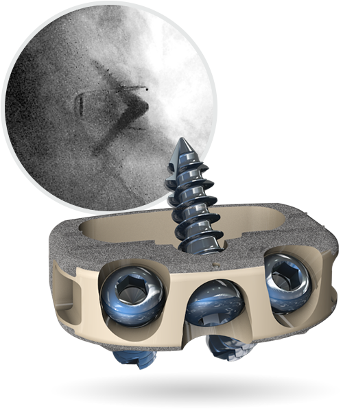
Blending the Benefits of Titanium and PEEK


By preserving its PEEK roots, STALIF M-Ti™ exhibits a modulus of elasticity similar to bone. In doing so, the device presents a reduced risk of subsidence relative to all-titanium cages.5 Additionally, STALIF M-Ti™ continues to maintain the radiolucent advantage of PEEK.


5 Ashman RB, et al. A continuous wave technique for the measurement of the elastic properties of cortical bone. J Biomech, 1984; 17(5):349-61.
6 Chen Y, Wang X, et al. Comparison of titanium and polyetheretherketone (PEEK) cages in the surgical treatment of multilevel cervical spondylotic myelopathy: A prospective, randomized, control study with over 7-year follow-up. Eur Spine J, 2013; http://dx.doi.org/10.1007/s00586-013-2772-y.
7 Centinel Spine Report VAL-2014-009.

Top of the Page ↑
Inherently STALIF®.
Standing on the Solid STALIF® Platform


STALIF M-Ti™ is built upon the simple, elegant design and proven success of the STALIF® foundation.

No-Profile®; No Supplemental Fixation

STALIF M-Ti's integrated cage and screw system is cleared for use without supplemental fixation, while the No-Profile® design allows it to nest fully within the confines of the vertebral body—leaving the anatomy unchanged external to the interbody.

Learn More

The Lag Effect

Compressive lag fixation ensures biomechanical stability and provides secure graft-endplate contact. Lag screws reduce the vertebral bodies onto the PEEK Cage creating graft / endplate surface compression to provide the best opportunity for fusion.

Biomechanical Equivalence

The STALIF M-Ti device is engineered to achieve equivalent biomechanical performance to anterior plate & cage, as well as posterior pedicle screw and cage, constructs.8
8 Cappuccino A, et al. Multi-Directional Flexibility Properties of the STALIF MIDLINE(tm) Device. October 2011.

Anti Back-Out® Screws

STALIF M-Ti™ screws provide an added layer of security with an Anti Back-Out® (ABO®) titanium split-ring which first compresses and then deploys during insertion.

For ordering information, please contact us at (484) 887-8810 or info@centinelspine.com


Our Products:

prodisc C
prodisc C Vivo
prodisc C SK
prodisc C Nova
prodisc L

The Story of Centinel Spine

Watch this three minute primer on Centinel Spine and its unique and extraordinary place as a catalyst of change in the spine industry—with pioneering technologies and a clinical history that have led to successes ranging from PGA champions to a growing list of surgeon-patients.


SEE MORE VIDEOS